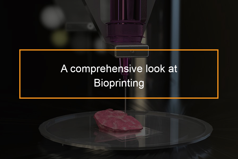
3D printing has advanced greatly recently and is permeating the health sector. The recent development of bioprinters that artificially construct living tissue by outputting layer-upon-layer of living cells is a marvel. Currently, all bioprinters are experimental. However, in the future, bioprinters could change medical practice.
What Is Bioprinting?
The basics of Bioprinting
Bioprinting is a type of 3D printing that uses cells and other biological materials as “inks” to fabricate 3D biological structures. Bioprinted materials can fix damaged organs, cells, and tissues in the body. Pretty soon, bioprinting may be used to build whole organs from scratch, a possibility that could transform the field of bioprinting and medicine in general.
The history of bioprinting
From its inception in the 80s to its use today in changing lives, 3D printing has come a long way. Charles Hull invented the first 3D printer 1984 that allowed tangible 3d objects to be made from digital data. The process has improved vastly in areas such as speed, material usage, and precision since its inception. In the early 1990s, MIT developed a procedure it trademarked with the name 3D Printing, formally abbreviated as 3DP. As of February 2011, MIT has approved licenses to 6 companies to use and promote the 3DP process in its products. While it previously took weeks to develop a 3D design, it now only takes a couple of hours. In 2000, Dr. Thomas Boland utilized a customized regular inkjet printer to print cells into a petri dish. In 2002, a miniature human kidney was developed at Wake Forest Institute for Regenerative Medicine.
In 2008, Objet Geometries Ltd. revealed its creation of a machine capable of printing one item utilizing a variety of products. This capability is important for bioprinting. Also in 2008, the first printed prosthetic leg was created and used. Australian automation specialist Invetech built the first 3D printer capable of making human organs and tissues, particularly for its partner Organovo.
In 2009, Organovo Inc. printed a capillary. In 2013, scientists at Heriot-Watt University 3D printed an arrangement of human embryonic stem cells.
Materials that can be used in bioprinting
Researchers have examined the bioprinting of many different cell types, which include stem cells, muscle cells, and endothelial cells. Several elements determine whether or not a material can be bioprinted. Initially, the biological materials had to be biocompatible with the materials in the ink and the printer itself. Also, the mechanical properties of the printed structure, in addition to the time it takes for the organ or tissue to develop, also affect the procedure.
Bioinks generally fall into one of 2 types:
Water-based gels, or hydrogels, function as 3D structures in which cells can flourish. Hydrogels containing cells are printed into specified shapes, and the polymers in the hydrogels are connected or “crosslinked” so that the printed gel becomes more powerful. These polymers can be organic or synthetic, but ought to work with the cells. All of the cells should spontaneously fuse into tissues after printing.
How Bioprinting Works?
The procedure of bioprinting
The bioprinting process is quite similar to the 3D printing process.
Preprocessing
An anatomically correct model is created digitally similar to how the target tissue or organs look like. Exact spatial details about the 3D location of cells is digitally generated and used to direct the printheads throughout usage. Cells taken from a patient’s biopsy, consisting of stem cells, are put in a development medium, where they increase to form a bio-ink of spheroidal cell aggregates.
Processing
The bio-ink is used to fill customized print cartridges, which are made up of syringes fitted with long extrusion nozzles. A hydrogel, or water-based compound, is first transferred separately within the bag to function as a short-lived mold and support.
The printheads, driven by computer system software application, deposit patterned cell aggregates accurately in the layers within the hydrogel, constructing them up in between interspersed layers of the gel component. When the model is completed, and deposition has ceased, the hydrogel is discarded. The bio-ink spheroids fuse without external interference to form the desired three-dimensional structure.
Post Processing
The tissue is taken from the printer and positioned in a damp environment, where it is expected to grow. Accelerated tissue maturation is intensified by the necessary mechanical and chemical conditioning of the bioprinted product. The tissue or organ is now all set for use as a transplant or research material.
Kinds of Bioprinters
Just like other types of 3D printing, bio-inks can be printed in various methods. Each approach has its own distinct benefits and downsides.
Inkjet-based bioprinting
Similar to an office inkjet printer. When using an inkjet printer, ink is fired through numerous small nozzles onto the paper. This develops an image made from lots of beads that are so little, and they are not noticeable to the eye. Researchers have adjusted inkjet printing for bioprinting, consisting of techniques that utilize heat or vibration to press ink through the nozzles. These bioprinters are more economical than other methods but are limited to low-viscosity bioinks, which could, in turn, constrain the kinds of materials that can be printed.
Laser-assisted bioprinting
Utilizes a laser to move cells onto a surface with high accuracy. The laser warms up part of the section, developing an air pocket and displacing cells towards a surface area. Since this technique does not need little nozzles like in inkjet-based bioprinting, greater viscosity materials, which can not flow easily through nozzles, can be used. Laser-assisted bioprinting enables extremely high accuracy when printing. However, the heat from the laser can damage the cells being printed. Moreover, the procedure can not quickly be “scaled up” to rapidly print structures in large amounts.
Extrusion-based bioprinting
Uses pressure to exact material out of a nozzle to create set shapes. This approach is reasonably versatile: biomaterials with different viscosities can be printed by changing the pressure, though care ought to be taken as higher pressures could probably damage the cells. Extrusion-based bioprinting can likely be scaled up for manufacturing; however, it may not be as precise as other methods.
Electrospray and electrospinning bioprinters
Make use of electrical fields to create droplets or fibers, respectively. These methods can have up to nanometer-level accuracy. Nevertheless, they utilize very high voltages, which might be unsafe for cells.
Applications of Bioprinting
Bioprinting Uses
Because bioprinting makes it possible for the exact construction of biological structures, the procedure may find a lot of application in medicine. Scientists have used bioprinting to mimic cells to help repair the heart after a cardiovascular disease along with depositing cells into injured skin or cartilage. Bioprinting has been used to produce heart valves for possible use in clients with cardiovascular diseases, build muscle and bone tissues, and aid in the repair of damaged nerves. Though more needs to be done to identify how these samples would perform in a medical setting, research studies show that bioprinting might be used to help regrow tissues during surgery or after injury. Bioprinters could enable the printing of entire organs, like livers or hearts, from scratch and used in organ transplants.
Uses of bioprinting
Skull Implants
The first 3D printed skull implant procedure was carried out on a 22-year-old female with an uncommon disorder in which the bone in her skull grew to nearly five times its initial thickness. Because of her condition, she had severe headaches and finally lost her eyesight. Three months after the 23-hour surgery, the client had made a complete recovery.
Facial Implants
The first 3D printed facial implant operation was carried out on a 29-year-old guy who wrecked his bike and even though he had a helmet, still damaged his cheekbones, shattered his eye sockets, damaged his nose and upper jaw, and fractured his skull. After modeling a 3D image of his skull utilizing CT scan technology, the doctors printed out the implants to rearrange the broken bones. The surgery was completed successfully, and the medical professionals intend to use the innovation in the future consistently.
Skin Implants
Wake Forest University is presently developing technology that sprays brand-new skin cells on burn wounds. First, a laser scans the injury and produces a brand-new, individualized strategy based on the width and depth of the injury. It then sprays brand-new skin cells, layer by layer, onto the burn until it is covered. This technique is superior to traditional skin grafts because it does not need the removal of skin from the patient, specifically when there is currently very little skin remaining. This innovation is presently being tested on mice, who healed two weeks quicker when treated like this.
Ear Implants
Cornell bioengineers and physicians have modeled an ear from a digital picture of a human ear and using 3D printing innovations to transform the image into a mold. They inject the mold with a combination of collagen from rat tails and 250 million cartilage cells from cow ears. After about a week, the ear is ready to be implanted on a person. Although they have effectively made human ear implants, tests still have to be run before they can be used, but Cornell biologists expect to be able to implant ears within three years.
Pelvic Implants
A 3D printed pelvis was put into a 60-year-old U.K. guy who contracted a rare kind of bone cancer known as chondrosarcoma. Orthopedic surgeons made use of a 3D printer to make a pelvis out of titanium powder, totally merged by a laser, which was then coated in “artificial bone” to allow for brand-new bone growth. After removing part of the patient’s hips to stop the cancer from spreading, the surgeons implanted the bioprinted hips. The man still needs to walk while using a cane. However, his implant was a complete success.
Prosthetic Jaws
The first 3D jaw implant surgery was carried out on an 83-year-old Belgian female with a persistent infection that eroded her jaw and justify her mandible . To breathe, eat, or perhaps talk, her jaw had to be removed. As a replacement, a 3D mandible was made, polished, and coated in “synthetic bone,” and implanted in the patient. She was given antibiotics to prevent infection, and soon her mouth was back to functioning as it should.
4D Bioprinting
In addition to 3D bioprinting, some groups have also analyzed 4D bioprinting, which takes into consideration the fourth dimension of time. 4D bioprinting is based on the concept that the printed 3D structures may continue to evolve gradually, even after they have been printed. The structures may thus change their shape and/or function when exposed to the right stimulus, like heat. 4D bioprinting may find use in biomedical fields, such as making capillaries by taking advantage of how some biological constructs fold and roll.
The pros and cons of bioprinting
3D bioprinting has a lot of benefits as well as some drawbacks.
Advocates of the innovation endorse the performance and ease of the printing process, while others condemn the printers as morally provocative and unattainable to the general masses.
Pros.
- Faster and more exact than standard techniques of rebuilding organs by hand.
- Less susceptible to human mistake.
- Less labor intensive for scientists.
- Organs are not likely to be rejected after transplantation.
- Lowers the occurrence of organ trafficking.
- Reduced waiting times for organ donors.
- Reduced animal testing.
- Finished parts are independent of biomaterial or scaffolding absent in native tissues.
- Effects of illness or drugs may be accurately observed without the need for human subjects.
- Reproduction of tissue is guaranteed through tight control of both composition and geometry and minimized variability.
- Efficient, diverse cell types permit improvement of tissue-specific functions.
Cons.
- Questions of liability if a printed object stops working.
- Contested ownership of the digital codes and implants produced.
- Numerous ethical concerns.
- Prices; it is quite expensive accessible only to the rich.
- It consumes a lot of energy.
- Emission of unhealthy particles into the air.
- A problem in keeping the cell environment, resulting in the death of numerous cells.
Facts about bioprinting
To help clear things up, we’ve made a list of 10 things to get you up to speed and at least help you figure out how bioprinting works and where it is headed in the future.
CT scans can operate like a CAD design
Instead of attempting to create an organ or tissue design from scratch, researchers and engineers can use a CT or MRI scan to create a 3D model to print. For instance, the University of Louisville, when developing a 3D printed model of a young boy’s heart so doctors might use it for his surgery, the scientists used the CT scan from his medical team to make the 3D model. Websites like Instructables even have tutorials to explain how to turn a CT scan into a 3D model to print.
There are several kinds of printers
Bioprinters: Organovo made the very first commercial bioprinter, called NovoGen MMX, which is the world’s very first production 3D bioprinter. The printer has two robotic print heads. One holds human cells, and the other holds a hydrogel, scaffold, or another type of material needed.
” Inkjet” influenced printers
Experiments with bioprinting at Wake Forest University were inspired by standard inkjet printers. The printer allows several cell types and components to be used for printing. In early prototypes, the cells were positioned in the actual walls of ink cartridges, and the printers were configured to place the cells in a specific order. Today, the university has modified that innovation so that skin cells can be positioned in an ink cartridge and printed straight on a wound.
The Six-axis printer
At the University of Louisville’s Cardiovascular Innovation Institute, Dr. Stuart Williams is Using a printer that, instead of constructing the tissue from the ground up, as standard 3D printers do, can build several parts of the heart tissue at the same time and move them around appropriately. ” We’ve made a six-axis printer that can print layers and come back and begin printing a brand-new layer on the outside of the heart,” Williams stated. “The valves are on one side, and we use robotics to bring the valves in and put them in parts of the heart.”.
Cells are used like “ink”
Generally, once a tissue design is picked, the manufacturer makes “bio-ink” from the cells. Using a bioprinter, the cells are layered in between water-based layers till the tissue is built. That hydrogel in between layers is often used to fill areas in the tissue or as support for the 3D printed tissue. Collagen is another medium used to fuse the cells. This layer-by-layer method is extremely comparable to the regular 3D printing process, where items are formed from the ground up.
Stem cells are also used in bioprinting
Stem cells can adjust quickly to tissues, so they are a good choice for bioprinting various organs and bones. Researchers at Nottingham University in the UK are exploring building bone replacements coated with stem cells that become tissue over time. The researchers proposed the increase of stem cell use in the repair of complex tissues, like those that make up the heart or the liver. It’s tough to use stem cells to build these organs, however, it may be possible with 3D bioprinting.
Bioprinting is more complex than other 3D printing
When it comes to Organovo, a bioprinter is used to create liver tissue, which is among the first experiments in bioprinting by the company. Spheroids of parenchymal (or essential) liver cells are packed into a syringe. In another one, nonparenchymal liver cells and the hydrogel, which merges to create a bio-ink, is filled. The bio-ink makes a mold in the petri dish, and the liver cells fill the rest of the dish. When the cells are incubated, they fuse to form the whole liver tissue.
There are numerous other materials to use in bioprinting
Cells don’t have to be the only material used in bioprinting. Lots of people still consider naturally degradable or biocompatible products that can be used to build body parts or repair injured ones as an element of bioprinting. Printing products that can enhance bones, cartilage, and skin are simply as crucial for the future of this innovation. Some of the products include specific types of flexible plastic, like the absorbable one used to make 3D printed windpipe splints for a child who had a condition that triggered his trachea to fall and titanium powder, which was used to create implants for a female who had an infection.
3D printed tissues for pharmaceutical testing
3D technology is not advanced to create a full organ, the tissue samples are best to test drugs and other medical advancements. Instead of using people or animals as guinea pigs for pharmaceutical testing, bioprinting might offer a more cost-effective and morally ethical option, while still being accurate because the tissue samples are made from human cells.
Replicating cells is nothing new
For years, researchers have been growing cells in laboratories, including skin tissue, blood vessels, and other cell cultures from various organs. Duplicating and growing cells in petri dishes is nothing new, and the science surrounding this is constantly improving. However, 3D printing presents an opportunity to print a whole organ, not just pieces of one.
Printing circulatory vessels is still a challenge
Vascularization is a big challenge in the printing of 3D organs because they need to have a system of arteries, blood vessels, and veins that support the system. They should be present to provide nutrients and get rid of waste created by the cells. One option is to leave the area in the 3D printed tissue for veins to be included later, but scientists are now attempting to determine a method to print blood vessels also.
One experiment at the University of Pennsylvania, a RepRap printer to make design templates of blood vessel networks out of sugar. When they dissolve, the sugar was rinsed without hurting the cells, and the space for the blood vessels exists. Scientists at Harvard have also begun dealing with this issue; however, they are attempting to 3D print the blood vessels by integrating them with skin cells.
The body can reject the 3D printed tissue
In any transplant or surgical treatment, there is constant worry of the body rejecting the organ or cells. This can even take place when tissue from one location of the body is taken into another area of the body. The organ (or piece of tissue) must have enough time to assimilate into the body after the implant. Since the technology for 3D bioprinting is still new, physicians and engineers have not even gotten to this part yet, however, it’s important to acknowledge these dangers ahead of time.
Bioprinting ethics
As with technology on the verge of innovation, 3D bioprinting has its own set of ethical problems.
One of the first problems is that of “playing God,” a term some use to express their unease with using this technology to outsmart fate or overlook “the will of God.” This perspective on biotechnology has caused a rift in its reception and therefore in the scope of its impact. This issue is closely associated with the possibility of immortality. The accessibility of bioprinting technology is at this point if human immortality is achieved through the procedure, it is most likely to be readily available to the wealthy. Earth’s human population would also increase, a value that some claim already goes beyond the planet’s carrying capacity.
Also, there is the possibility of strengthened “cyborg” organs, printed using non-human components. This idea agitates the problems above, also conjuring pictures of a more robotic society. The concern of quality assurance also exists. Until now, there are no steps in place to guarantee that bioprinted organs are of excellent health legally. This lack of policy might lead to the exploitation of unknowing patrons in the creation of unhealthy or low-quality body parts.
The Future of bioprinting
The future of bioprinting is promising. Scientists at Wake Forest are presently testing makers that can print skin directly onto a patient’s body, a procedure to be used on burn and trauma victims. Although 3D printers are presently a high-end, their costs have reduced significantly; initially priced at around $10,000, 2014 saw the development of the $100 Peachy Printer.
Miniature 3D printed organs called “organoids” are presently being used to evaluate the reaction of human organs to conditions otherwise impossible or dangerous to recreate in people. Physicians have been scanning and 3D printing plastic models of patients’ hearts, allowing them to see its problems before choosing real surgical treatment. A cardiologist at the Children’s Hospital of Illinois is trying to construct a catalog on the study of these hearts so that medical students and doctors can quickly access it for their research studies.
A professor of bioengineering, Paul Calvert, Ph.D., at the University of Massachusetts at Dartmouth, sees the capacity for bioprinting complete, full-sized organs later on, however, he notes that “laboratory samples for screening, diagnosis, and tailored production of active proteins have to be more detailed.”.
If 3D bioprinting technology becomes as widespread as researchers are envisioning, operators will be able to order tailored organs. This would significantly enhance the current system of waiting on a donated organ for transplanting. If we can use 3D bioprinting for customized medicine, there is the possibility to achieve immortality. After all, what is stopping us from simply printing new body parts when ours begin to age and stop working?


